Richard Suter
Richard Suter trained as a school teacher before beginning to prepare and sell microscope slides in the late 1880s, running his enterprise from the family home on Highweek Road, Tottenham. By 1892, his advertisements announced “50,000 choicest microscopic slides of every description in stock.” His slides are identified by their distinctive pink or salmon labels, hand-lettered in his characteristic script. The label on the blackprint slide reads: “Exhibition Slide. Polycystina from Barbados. Richard Suter, 10 Highweek Road, Tottenham.” An exhibition slide was a higher-order preparation — arranged for visual impact as much as for scientific study, intended to demonstrate the maker’s skill at its best.
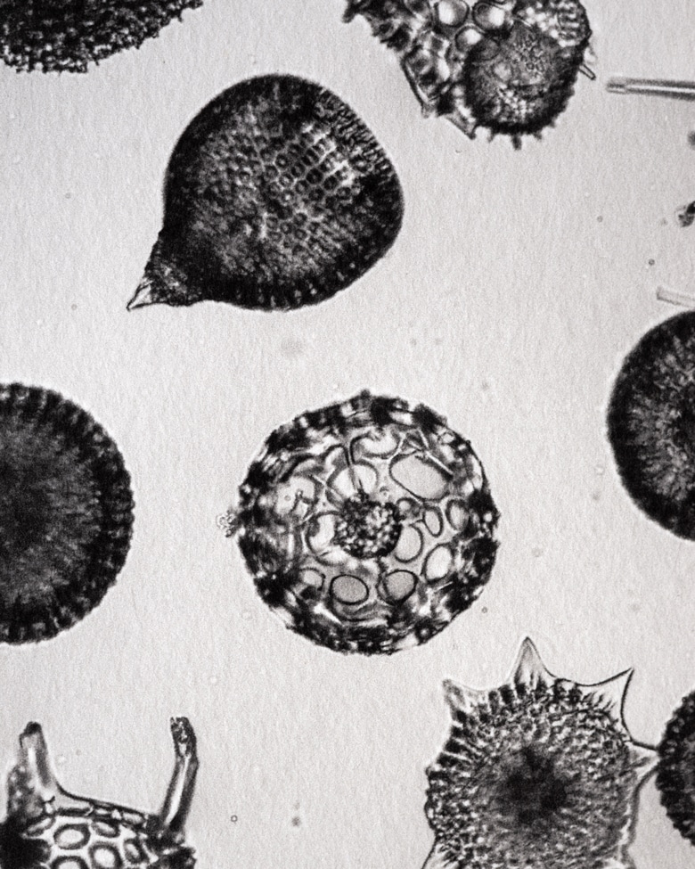
The Polycystina of Barbados
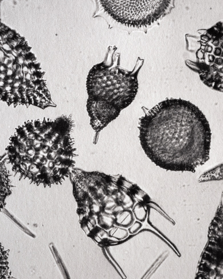
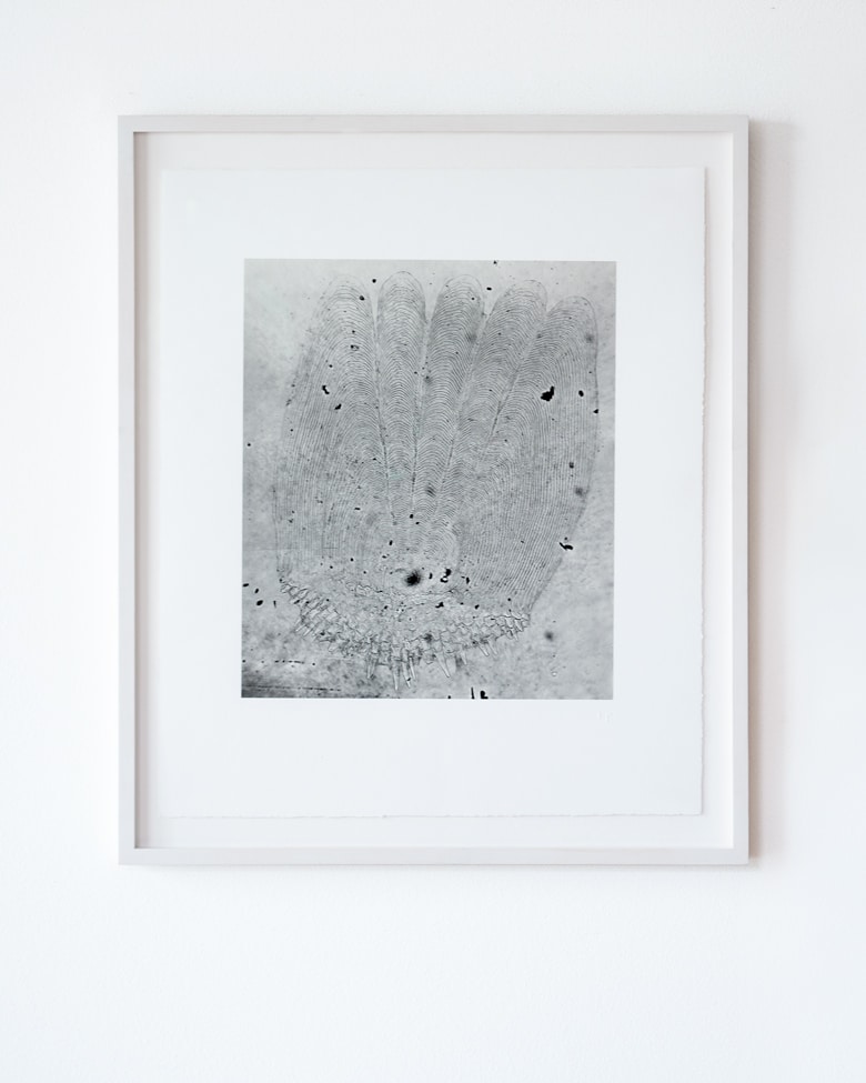
Polycystina are radiolarians: single-celled marine organisms that secrete intricate silica skeletons — spherical, latticed, spiked — of a complexity that bears no relation to their scale. Individual skeletons range from roughly 50 to 500 micrometres. In 1846, the physician John Davy discovered a substantial deposit of polycystine skeletal remains in a sedimentary layer on the Caribbean island of Barbados, and brought samples back to England. The deposit is Eocene in origin — the remains of organisms that lived between approximately 35 and 45 million years ago, accumulated on an ancient ocean floor and later lifted to the surface through the tectonic processes that formed the island. The material circulated rapidly among microscopists, who found that the Barbados earth — treated with acid to dissolve the surrounding carbonate — yielded slides of exceptional diversity and clarity. Suter’s preparations were among the most sought-after objects in the Victorian slide trade. What he arranged on his slide are not fossils in the conventional sense: not mineral casts that replaced an original structure, but the original silica skeletons produced by the organisms themselves, stable enough to survive intact for tens of millions of years. The objects in the print are the objects that were alive.
The reconstruction
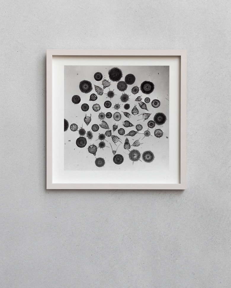
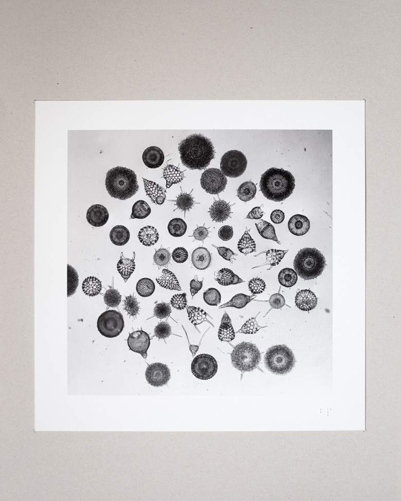
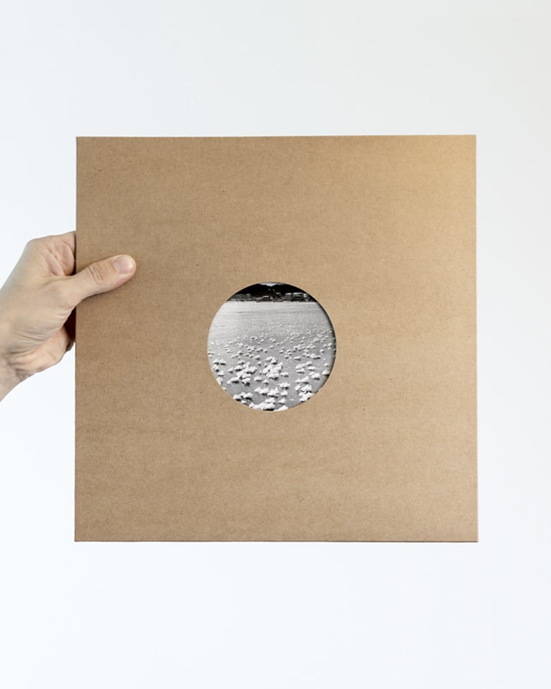
The original grouping of polycystine skeletons assembled by Suter on his slide measures 2mm across. A microscope lens operates at extremely short focal depth: at the magnification required to resolve the surface structure of individual skeletons, only a fraction of a millimetre lies in focus at any one time. To reconstruct the full volume of Suter’s arrangement, approximately one hundred photographs were made at successive focal planes, then combined into a single composite image. Printed at 30 × 30 cm on Arches Velin BFK Rives, the skeletons appear at roughly 150 times their actual size — at a scale at which their architectural complexity becomes the primary fact of the image.
The plate
Microscope slide, 2.6 × 7.5 cm, blackprint collection. Prepared by Richard Suter, 10 Highweek Road, Tottenham, England. Label: “Exhibition Slide. Polycystina from Barbados.”