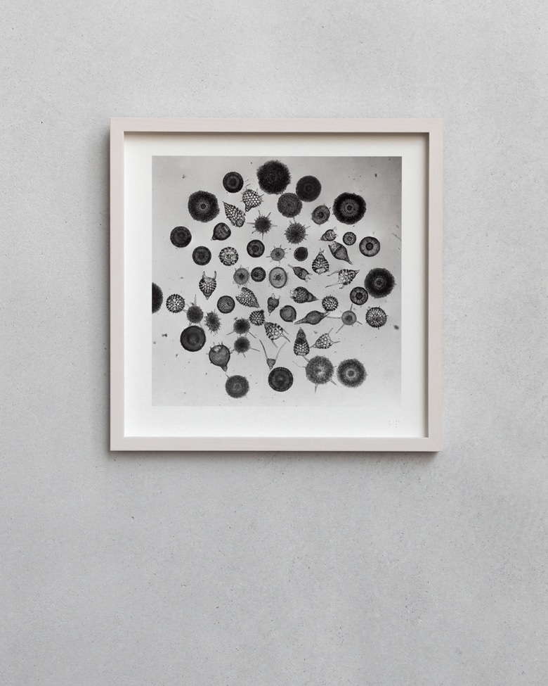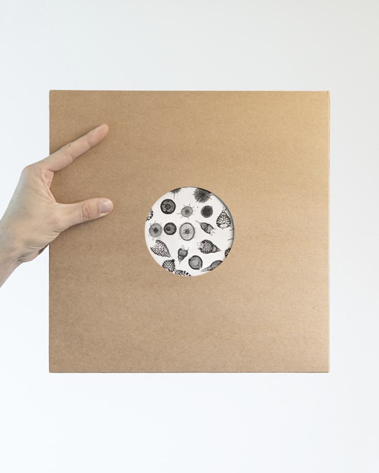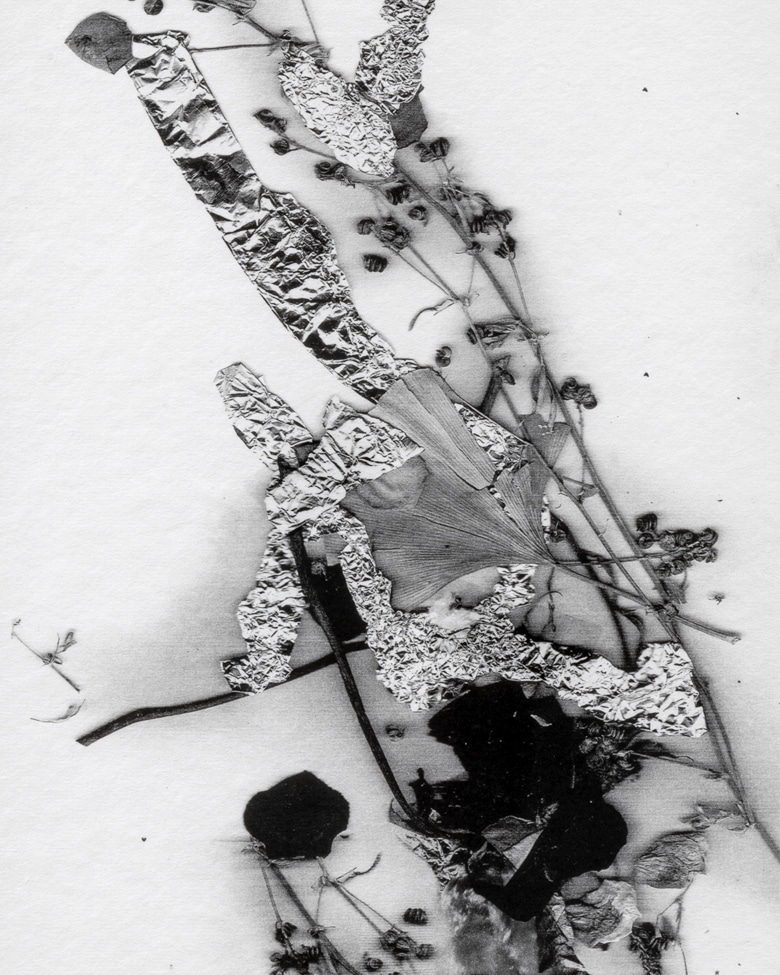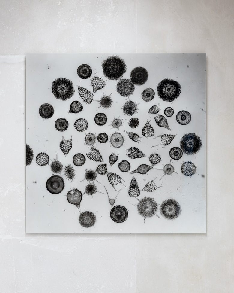

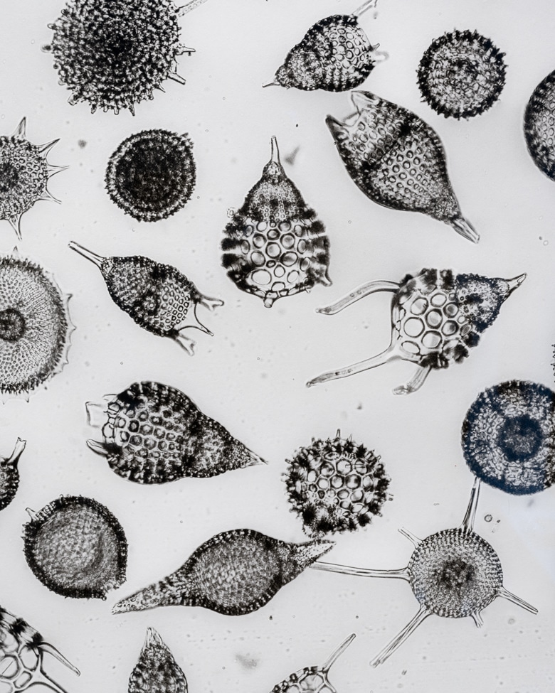
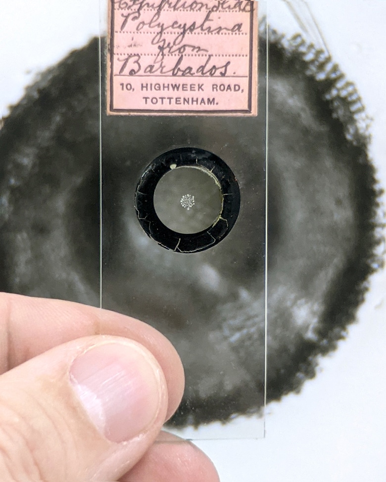

Title: “Polycystina from Barbados”
Year: 1890
Country: England
Photographer: Richard Suter
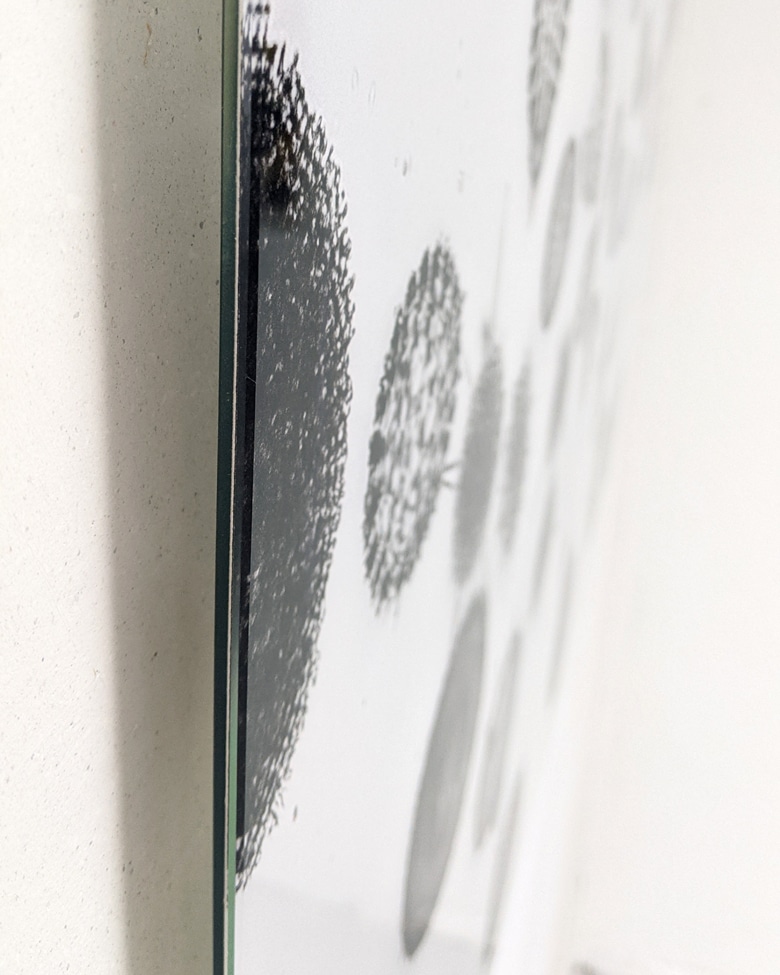
Origin: Microscope slide 2.6 × 7.5 cm / The blackprint collection
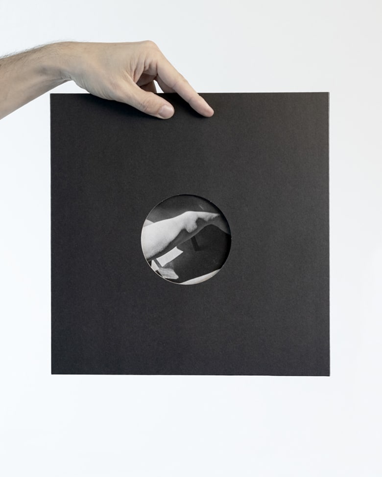
Size Glass & Print: 89.5 × 89.5 cm | 89.5 × 89.5 cm
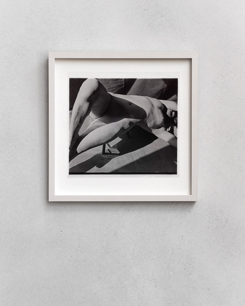
Printing technique: Piezography® Pro
Paper: A.I.J.P Awagami Bamboo 170 gr/m2. pH-neutral.
Laminated Glass: Front: HY-TECH-GLASS, 2 mm non-reflective extra-clear. | Back: Glass, 2 mm float.
Weight: 8 Kg
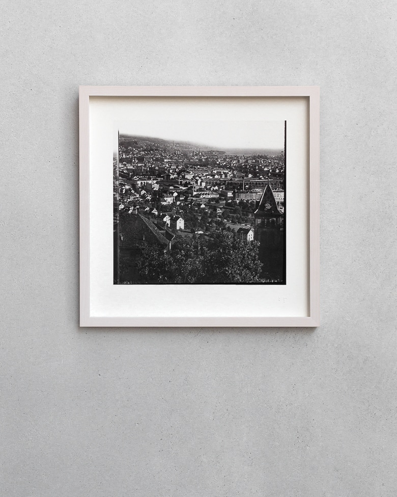
First edition of 15, Zürich
blackprint with Abgraphics Studio
Provided with a Verisart blockchain Certificate of Authenticity.